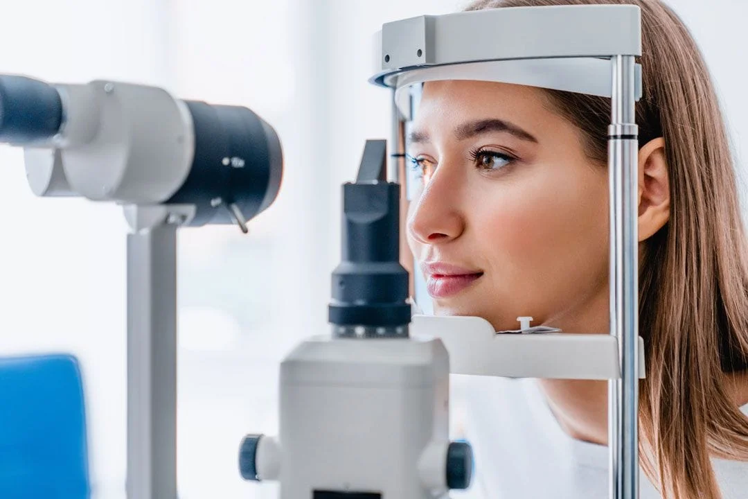
Zeiss Clarus 500 Retinal Imaging
Clarus 500 is the newest ultra-widefield retinal camera from Zeiss, that aids our optometrists in the diagnosis and documentation of ocular disease. In addition to true color imaging, it also captures high-resolution fundus autofluorescence images and external eye images. It is a touch-free, painless scan that takes only seconds to complete. The images these scans produce can help our doctors identify signs of vision-threatening diseases such as glaucoma, macular degeneration, and diabetic retinopathy. It allows our optometrists to track subtle changes in pathology over time. Therefore, we recommend a baseline retinal screening image for all of our patients as part of their annual health examination.
Zeiss Humphrey Field Analyzer
A visual field test maps measures the entire area of peripheral vision that can be seen while the eye is focused on a central point.. The visual field shows changes that are not noticed by the patient until damage is severe. The visual field can detect damage caused by diseases, such as glaucoma, stroke, macular degeneration and diabetes. Usually, the visual field test is taken once a year but depending on the severity of your eye disease, your optometrist may decide to check your visual field more frequently.
Zeiss Cirrus HD-Optical Coherence Tomography (OCT)
Optical coherence tomography (OCT) is a non-invasive imaging test which uses light waves to take cross-section pictures of your retina, so our optometrists can see each of the retina’s distinctive layers. Mapping and measuring the thickness of each layer helps our doctors with diagnosis and treatment guidance for glaucoma and other disease of the retina, such as age-related macular degeneration, macular edema and macular holes
Marco OPD Scan III Corneal Topographer
Corneal topography maps the surface of your cornea, similar to a topographic map. It shows distortions in the curvature of the cornea, raised surfaces and other surface irregularities. It allows our optometrists to discern conditions which are otherwise undetectable to conventional testing methods. This equipment is highly effective in the diagnosis and treatment of multiple conditions, including astigmatism, keratoconus, corneal dystrophies, and corneal scars or opacities.
